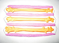

I began by forming the bone structure. First is detail of the ball and socket joint that connects the pectoral (shoulder) girdle and the upper limb.
The large bone at the top of the arm is the humerus. This is joined to the two lower bones in the arm: the radius and the ulna. (The humerus is connected to the ulna with a hinge joint).

Next is detail of the arm at rest.
The muscles here are represented by the pink playdough.
Tendons are represented by the blue. Tendons enable the muscle to move the bone.
In the first image below, the bicep muscle is relaxed, while the tricep muscle is contracted.
In the second image the opposite is true. (This is because the bicep and the tricep muscles are antagonistic--meaning that they work in opposition).


Prior to the change in arm position, a neuron would carry a message to the muscle, signaling the muscle to contract. The image below shows a neuron signaling the bicep to contract.

A neuron is represented by orange colored playdough. The signal being sent by the neuron is also known as a nerve impulse or action potential. The Schwann cell (and myelin sheath) are represented by the smallest blue pieces of playdough within the orange (*not to be confused with the blue tendons--I ran out of colors!).
Ugh, I can not get these pictures to format. From left to right they are A, B, and C.A
D
Image A shows a motor neuron. Again, Schwann cells are represented by blue. Neurotransmitters are located at the end of the strands in axon terminals. These send messages between the neurons.
Image B shows sarcomeres that are contracted. Image C shows sarcomeres that are relaxed. Both images represent the sliding filament process. Actin is represented by the pink, while myosin is represented by the orange. During this process the sarcomere shortens and actin filaments slide past the myosin filaments.
Image D shows action potential. The nerve impulse passes/is sent rapidly from one section to another.
Thus, the entire systems works together to enable movement. Movement begins as the central nervous system transmits a message via neurons. Nerve impulses then initiate the action. In conclusion, the process of initiating human movement likely simulates the process of initiating movement of other organisms. (image)
(Also, an informative page on the sliding-filament theory of muscle action can be found here).
*source: Mader, Sylvia. Human Biology, tenth edition.


1 comment:
Stephanie Rose
COMPENDIUM MAJOR TOPIC ONE: NERVOUS FUNCTION:
Beautiful complete compendiums with great images well incorporated…good info…sources all given…keep it up!
COMPENDIUM MAJOR TOPIC TWO: MOVEMENT:
LAB MAJOR TOPIC ONE: LEECH NEURONS:
Great job with good answers to questions
LAB MAJOR TOPIC TWO: MUSCLE FUNCTION:
Nice data and good explanations…yes, cycling of calcium and also other nutrients the cells need probably are slower in cold.
LAB PROJECT: LIMB MODEL:
Great limb…clearly shows all basic elements..I’m not sure we can exactly see how muscle actin-myosin contraction will cause movement in limb…but more on this in A and P
ETHICAL ISSUE ESSAY: ACTIVITY AND EXERCISE:
I think you’re right—lack of time and money—do we need to pay people to get active!
Stephanie—great job on this basically perfect unit! The compendiums are beautiful—keep it up!
LF
Post a Comment