
Chapters Thirteen and Fourteen: Nervous Function
(image source)
What does a nervous system do? (page 270, web)
Diffusion and Action Potentials in Neurons-- (online lab link, Powerpoint, page 270)
rapid transmission of messages (Powerpoint)
Reflex arc (simple somatic function) (Wikipedia)
autonomic function (Powerpoint, page 270)
What can we sense? (pages 275-293)
The nervous system is divided into the central nervous system and the peripheral nervous system. The purpose of the system is to receive input, to integrate data, and to generate motor output. Nervous tissue consists of two types of cells. These are neurons and neurolgia. Neurons transmit nerve impulses, while neurolgia nourish and support them. Sensory neurons take nerves impulses from sensory neurons to the central nervous system. Motor neurons take nerve impulsed away from the central nervous system to muscles or glands.
The central nervous system consists of the spinal cord and brain.
 The image to the left shows that different levels of the spinal cord control different parts of the body. The spinal cord is a column of nervous tissue that acts as a pathway to transmit information from the brain to the body.
(http://www.reeve.uci.edu/anatomy/images/scns_1b.gif)
 The image to the right shows the brain and its components. The brain has three main components: the cerebrum, cerebellum, and brain stem.
The cerebrum controls sensation, reasoning, learning, memory, language, and reasoning. The cerebellum coordinates skeletal muscle contractions. The brain stem mainly controls unconscious vital signs such as breathing and heartbeat.
The Peripheral Nervous System contains only nerves and ganglia. Cranial nerves take impulses to and from the brain, while spinal nerves take impulses to and from the spinal cord.
Resting and action potential are explained in great detail in the leech lab for this unit.
Another animation of the process can be seen here.
In resting potential there is more Na+ outside the axon and more K+ inside. (This does not conduct an impulse). In action potential a nerve impulse occurs. As Na+ gates open, Na+ moves to the inside of the axon, resulting in a depolarization. When the K+ gates opens and K+ moves to outside the axon, a repolarization occurs.
Somatic System The somatic system serves the skin, skeletal muscles, and tendons. Some actions are due to reflexes which are automatic and involuntary. Others are voluntary and originate in the cerebral cortex.
"A reflex arc is an automatic reaction that allows an organism to protect itself reflexively when an imminent danger is perceived. In response to certain stimuli, such as touching a hot surface, these reflexes are 'hard wired' through the spinal cord. A reflexive impulse travels up afferent nerves, through a spinal interneuron, and back down appropriate efferent nerves". (websource: wikipedia).
Autonomic System The autonomic system is divided into two divisions: sympathetic and parasympathetic. The sympathetic division includes responses that occur during times of stress. The parasympathetic division consists of responses that occur during times of relaxation. Actions in these divisions are both automatic and involuntary. These divisions innervate internal organs. (Two neurons and one ganglion are used for each impulse).
Senses
There are four types of sensory receptors: chemoreceptors, photoreceptors, mechanoreceptors, and thermoreceptors. Sensory receptors initiate nerve impulses that are transmitted to the spinal cord/brain. Sensation occurs when nerve impulses reach the cerebral cortex. Perception is an interpretation of sensations.
A. Proprioceptors
1. mechanoreceptors involved in reflex actions
2. help maintain equilibrium and posture
B. Cutaneous Receptors
1. found in skin
2. for touch, pressure, temperature, and pain
The senses of taste and smell are due to chemoreceptors that are stimulated by molecules in the environment. This site has a really interesting article relating to the taste buds and different senses of taste. (bitter, sour, sweet, salty).

(Microvilli of taste cells have receptor proteins for molecules that cause the brain to distinguish between the tastes).
 The cilia of olfactory cells have receptor proteins for molecules that cause the brain to distinguish odors.
The sense of vision depends on the eye, the optic nerves, and the visual areas of the cerebral cortex.

![]() The eye has three layers. The sclera (or outer layer) protects and supports the eyeball.
The eye has three layers. The sclera (or outer layer) protects and supports the eyeball.
The choroid (middle, pigmented layers) absorbs stray light rays.
The retina (inner layer) contains the rod cells (sensory receptors for dim light) and cone cells (for bright light and color).
The lens (along with cornea and humors) brings the light rays to focus on the retina. To see a close object a visual accommodation occurs as the lens rounds up.
The visual pathway begins when the light strikes photorecptors (rod cells and cone cells) in the retina. The optic nerves carry nerve impulses from the eyes to the optic chiasma, then pass through the thalamus before reaching in primary vision area. (the occipital lobe of the brain).
Hearing depends on the ear, the cochlear nerve, and the auditory areas of the cerebral cortex.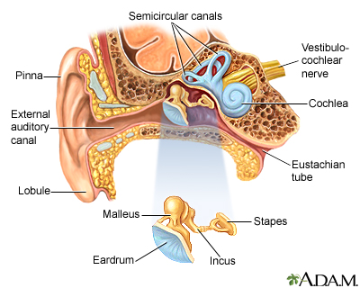
The ear has three parts. In the outer ear, the pinna and the auditory canal direct sound waves to the middle ear. In the middle ear the malleus, incus, and stapes amplify sound waves. In the inner ear, the semicircular canals detect rotational equilibrium.
The auditory pathway begins when the outer ear receives and the middle ear amplifies sound waves that then strike the oval window membrane.
The ear also contains mechanorecptors for equilibrium.

No comments:
Post a Comment