Chapters Eleven and Twelve
Muscle Cells: Contraction and Calcium (Powerpoint, web, pages 230-242)
Movement Across Joints (Powerpoint, web, page 224)
Bones, Bone Tissue, and Calcium (Powerpoint, web, pages 208-222)
Types of Muscle
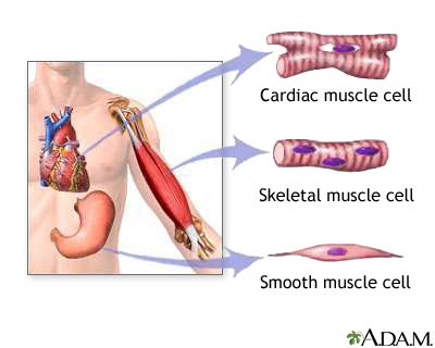
A. Smooth Muscle
1. involuntary
2. occurs in walls of internal organs
B. Cardiac Muscle
1. Involuntary
2. occurs in walls of heart
C. Skeletal Muscle
1. Voluntary
2. helps maintain posture
3. provides movement and heat
4. protects underlying organs
Muscle cells are pretty large--each containing hundreds of myofibrils. Myofibrils contain actin and myosin filaments. Muscle contraction occurs when sarcomeres shorten and actin filaments slide past myosin filaments.
Calcium Release in a Muscle
1. A motor neuron impulse arrives to axonal terminus
2. neuro-muscular junction synapse passes message along to muscle cell
3. muscle cell membrane undergoes action potential along length of cell membrane and into T-tublue system
4. voltage change causes release of calcium ions into muscle cell
5. calcium causes actin-myosin units to shorten
The shortening of actin-myosin units leads to whole muscle contraction.
Whole Muscle Contraction
A. Muscle cells all lined up in skeletal muscle
B. Contraction of many cells makes the whole muscle shorten, bringing about body movement
C. Movement happens across joints between skeletal elements
 Muscle contraction as described in detail from the above web source.
 Joints connect skeletal elements. Synovial joints are lubricated, mobile joints. Skeletal elements are linked by synovial joints and move when muscles pull on those skeletal elements.

The image above shows the different types of synovial joints.
Fibrous joins are immovable. (i.e. sutures of the cranium).
Cartilaginous joints are slightly moveable (such as those between the ribs and sternum and pubic symphysis).
A Bone is alive, has nerves, blood supply, and cells. Cells make bone tissue, dissolve bone tissue, and live inside bone tissue. Bone also serves as calcium storage. Calcium is needed for many aspects of cell metabolism, but especially in muscle cells.
Bones contain more calcium than any other organ. When blood calcium levels decrease below normal, calcium is released from the bones so that there will be an adequate supply for metabolic needs. When blood calcium levels are increased, the excess calcium is stored in the bone matrix. The dynamic process of releasing and storing calcium goes on almost continuously. (websource)

The image to the left is a diagram of bone cells.

Bone remodeling is the renewal of bone. Some bone is recycled each year to allow the body to regulate blood calcium.
Axial Skeleton
1. skull (formed by cranium to protect brain and facial bones)
2. hyoid bone (anchors tongue)
3. vertebral column (supports head/trunk, protects spinal cord, site for muscle attachment)
4. rib cage (protects heart, lungs)
Appendicular Skeleton
1. pectoral girdles (flexibility)
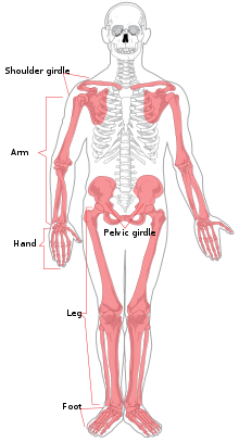 2. upper limbs (flexibility)
3. pelvic girdles (supporting weight)
4. lower limbs (supporting weight)
There are two types of bone tissue: compact and spongy. The names imply that the two types of differ in density, or how tightly the tissue is packed together. There are three types of cells that contribute to bone homeostasis. Osteoblasts are bone-forming cell, osteoclasts resorb or break down bone, and osteocytes are mature bone cells. An equilibrium between osteoblasts and osteoclasts maintains bone tissue. (websource).
An interesting article on bone tissue transplantation can be found here.
The National Osteoporosis Foundation has a good website with tips for maintaining bone health.

1 comment:
thank you! Although I believe the image of the skeletal joints isn't functioning properly...
Post a Comment